מגזין
אסטקסנטין ובריאות העור, טיפול במחלות ועוד

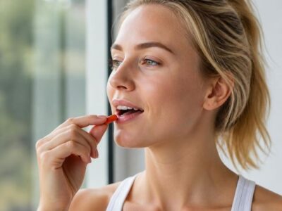
אסטקסנטין הינו קרוטנואיד, הוא מטבוליט משני המסונתז באופן טבעי על ידי מספר חיידקים, מיקרו-אצות ושמרים. הייצור המסחרי של פיגמנט זה בוצע באופן מסורתי על ידי סינתזה כימית, אך נראה שהמיקרו אצה Haematococcus pluvialis היא המקור המבטיח ביותר לייצור הביולוגי התעשייתי שלה. בשל הפונקציות המגוונות הקולקטיביות שלו בביולוגיה של העור, ישנן עדויות מתגברות לכך שאסטקסנטין הוא בעל יתרונות בריאותיים שונים ויישומים תזונתיים חשובים בתחום הדרמטולוגיה.
למרות שעדיין מתווכחים, הוצעו מגוון מנגנונים פוטנציאליים שבאמצעותם astaxanthin עשוי להפעיל את היתרונות שלו על הומאוסטזיס של העור, כולל השפעות פוטוגנטיות, נוגדות חמצון ואנטי דלקתיות. סקירה זו מסכמת את הנתונים הזמינים על התפקיד התפקודי של אסטקסנטין בפיזיולוגיה של העור, מתווה מנגנונים פוטנציאליים המעורבים בתגובה לאסטקסנטין, ומדגישה את ההשלכות הקליניות הפוטנציאליות הקשורות לצריכתו.
יתרונות האסטקסנטין
פעילות נוגדת חמצון:
לחץ חמצוני ממלא תפקיד מכריע בהזדקנות העור האנושי ובנזק העורי. המנגנונים של הזדקנות פנימית (כרונולוגית) וחיצונית (צילום) כוללים יצירת מיני חמצן תגובתיים (ROS) באמצעות חילוף חומרים חמצוני וחשיפה לאור אולטרה סגול (UV) של השמש, בהתאמה. לפיכך, היווצרות ROS היא מנגנון מרכזי המוביל להזדקנות העור. אירועי חמצון של הזדקנות העור כוללים נזק ל-DNA, תגובה דלקתית, ייצור מופחת של נוגדי חמצון, ויצירת מטריקס מטלופרוטאינזים (MMPs) המפרקים קולגן ואלסטין בשכבת העור העורית [16,17,18]. ישנם מקורות תזונתיים או אקסוגניים רבים הפועלים כנוגדי חמצון, כולל פוליפנולים וקרוטנואידים [19,20]. ASX תפס לאחרונה את התעניינותם של חוקרים בגלל פעילות נוגדת החמצון החזקה שלה ותכונות השליח המולקולריות והביוכימיות הייחודיות שלה עם השלכות בטיפול ומניעת מחלות עור. מחקרים השוואתיים שבדקו את ההשפעות הפוטו-פרוטקטיביות של קרוטנואידים הוכיחו ש-ASX הוא נוגד חמצון מעולה, בעל יכולת נוגדת חמצון גדולה יותר מאשר קנתקסנטין ו-β-קרוטן בפיברובלסטים עוריים אנושיים. בפרט, ASX מעכב היווצרות ROS ומווסת את הביטוי של אנזימים המגיבים ללחץ חמצוני כגון Heme oxygenase-1 (HO-1), שהוא סמן ללחץ חמצוני ומנגנון ויסות המעורב בהסתגלות התא נגד נזק חמצוני [21] ]. HO-1 מווסת באמצעות גורמי שעתוק שונים רגישים ללחץ, כולל גורם גרעיני קשור לגורם אריתרואיד 2 (Nrf2), אשר נקשר לאלמנטים של תגובה נוגדת חמצון באזורי הפרוטור של אנזימים של חילוף החומרים המבטל רעלים [22]. מספר מחברים הוכיחו ש-ASX מפעיל את מסלול נוגד החמצון Nrf2/HO-1 על ידי יצירת כמויות קטנות של ROS [23,24]. בהתאם למחקרים אלו, Xue et al. [25] ראה ש-ASX הגביר את ביטוי Nrf2 בתאים מוקרנים. יתר על כן, החלבונים ממוקדי Nrf2 HO-1 והאנזימים נוגדי החמצון סופרוקסיד דיסמוטאז 2 (SOD2), קטלאז (CAT) וגלוטתיון פרוקסידאז 1 (GPX1) הווסרו באופן משמעותי בתאים מוקרנים בנוכחות ASX. לכן, ASX מפעיל פעילויות נוגדות חמצון משמעותיות לא רק באמצעות ניקוי רדיקלי ישיר, אלא גם על ידי הפעלת מערכת ההגנה נוגדת החמצון התאית באמצעות אפנון של מסלול Nrf2. מחקר שנערך לאחרונה גם הראה ש-ASX הגן מפני התקדמות מוקדמת של פצעי כוויה על ידי החלשת מתח חמצוני המושרה על ידי ROS במודל של צריבה עמוקה של חולדה. השפעה זו כוללת ויסות של ייצור רדיקלים חופשיים על ידי השפעה על קסנטין אוקסידאז (XO) ועל הצורה המופחתת של ניקוטינמיד אדנין דינוקלאוטיד פוספט (NADPH) אוקסידאז (Nox); שניהם תורמים ליצירת ROS [26].
תכונות אנטי דלקתיות:
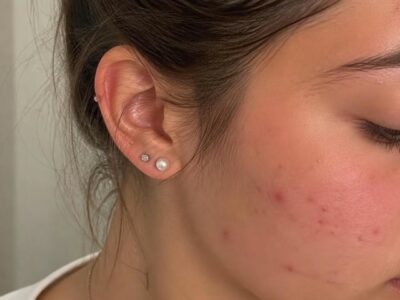
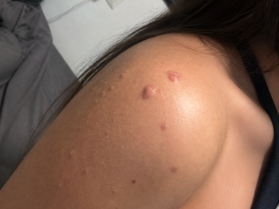
מחקר מקיף במהלך שני העשורים האחרונים חשף את המנגנון שבאמצעותו מתח חמצוני מתמשך מוביל לדלקת כרונית, אשר בתורה, מתווך את רוב המחלות הכרוניות כולל ניוון עצבי, סרטן ונזק לעור [27,28,29]. ידוע היטב כי סמנים פרו-דלקתיים שונים בעור גדלים כתוצאה מחשיפה ל-UV. לקרטינוציטים תפקיד מכריע בתגובת הפוטו-דאג’ לאחר חשיפה ל-UV על ידי שחרור מתווכים פרו-דלקתיים. הוכח שטיפול ב-ASX מונע את ההשפעות המזיקות של UV על ידי הפחתת ייצור מיני חנקן תגובתי המושרה על ידי UV, ביטוי ציטוקינים דלקתי ואפופטוזיס בקרטינוציטים. ASX גרם לירידה משמעותית ברמות של תחמוצת החנקן הניתנת לשרירה (iNOS) ו-cyclooxygenase (COX)-2, והפחיתה את שחרור הפרוסטגלנדין E2 מקרטינוציטים לאחר הקרנת UV [30]. להשפעה המעכבת של ASX על ייצור iNOS יש השלכות חשובות על פיתוח תרופות אנטי דלקתיות למחלות עור דלקתיות כמו פסוריאזיס ואטופיק דרמטיטיס (AD). AD היא מחלת עור דלקתית כרונית הקשורה לגורמים שונים, כולל הפרעות אימונולוגיות התורמות לפתוגנזה ולהתפתחות נגעים בעור. דוח שנערך לאחרונה הראה ש-ASX עיכב את ביטוי הגנים של מספר סמנים ביולוגיים פרו-דלקתיים כגון אינטרלוקין-1β (IL-1β), אינטרלוקין-6 (IL-6) ו-Tumor necrosis factor-α (TNF-α) במודל חיה AD [31]. מספר חוקרים בחנו את העיכוב של גורם גרעיני-קאפה B (NF-κB) על ידי ASX. בפרט, דווח כי ל-ASX יש יכולת חזקה לחסום את הטרנסלוקציה הגרעינית של תת-היחידה NF-κB p65 ופירוק IκBα באמצעות השפעתה המעכבת על פעילות NκB קינאז (IKK) [32]. חשוב מכך, מחקרים הראו את היכולת של ASX לעכב את הייצור של מתווכים דלקתיים על ידי חסימת הפעלת NF-κB בקרטינוציטים אנושיים, מה שמצביע על כך ש-ASX עשויה להציע אסטרטגיה חדשה ומושכת לטיפול במחלות דלקתיות בעור [33].
השפעות משפרות את מערכת החיסון:
ראיות ניכרות מצביעות על כך שדיכוי המערכת החיסונית תורם להתפתחות של ממאירות עורית הנגרמת על ידי UV, כולל מלנומה ולא מלנומה, הן במודלים של עכברים והן בבני אדם [34,35,36]. ASX משפיע באופן משמעותי על תפקוד מערכת החיסון במספר מבחני in vitro ו-in vivo [37]. לדוגמה, מחקרים במבחנה על לימפוציטים אנושיים הוכיחו שיפור על ידי ASX של ייצור אימונוגלובולינים בתגובה לגירויים תלויי תאי T [38]. הפעולה האימונומודולטורית של ASX דווחה גם בכלבים וחתולים, מה שמגביר את התגובה החיסונית בתיווך התא וגם בהומור. במחקרים אלו, ASX הגביר את הפעילות הציטוטוקסית של תאי הרוצח הטבעי (NK), דבר המצביע על כך ש-ASX עשוי לווסת תאי NK המשמשים כמערכת מעקב חיסונית כנגד גידולים ותאים נגועים בנגיף [39,40]. יתרה מכך, מחברים אחרים הראו ש-ASX הגביר את פעילות לימפוציטי T ציטוטוקסיים בעכברים. תאי T פעילים ותאי NK מייצרים אינטרפרון-γ (IFN-γ), המעורב בוויסות החיסון ובהתמיינות תאי B; לפיכך, ASX עשוי להגביר את התגובות החיסוניות ועלול להפעיל פעילות אנטי-גידולית [41]. בנוסף לתגובה החיסונית בתיווך התא, כפי שכבר הוזכר, ASX גם עורר חסינות הומורלית. ASX הגביר את ייצור הנוגדנים בטפלנוציטים של עכברים, החזיר את התגובה החיסונית ההומורלית בעכברים ישנים, וגרם לייצור של נוגדנים רב-שבטיים G ו-M בתאי טחול עכברים [42,43,44]. למרות שדרושים מחקרים נוספים כדי להבהיר טוב יותר את אופן הפעולה הספציפי של ASX בהגברת התגובה החיסונית, יחד, תצפיות אלו מצביעות על כך ש-ASX עשוי להיות כלי פוטנציאלי נגד דיכוי חיסוני המושרה על ידי UV.
השפעות על נזקי עור:
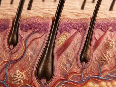
המבנים החשובים והנפוצים ביותר של המטריצה החוץ-תאית העורית (ECM) הם קולגן, אלסטין וגליקואמינוגליקנים (GAG). הן בהזדקנות הפנימית והן בהזדקנות החיצונית, נצפים שינויים במבנים אלה. שינויים אלו מובילים לאובדן חוזק מתיחה ויכולת רתיעה, היווצרות קמטים, יובש ופגיעה בריפוי פצעים [45]. בנוסף, ROS המושרה על ידי UV ממריץ את הסינתזה של MMPs שאחראים לפירוק של ECM, ובפרט, MMPs יכולים לפרק קולגן באופן מלא [46]. במבחנה, ASX מדכא ביעילות נזק לתאים הנגרם על ידי רדיקלים חופשיים והשראת MMP-1 בעור לאחר הקרנת UV [47]. כמה מחקרים דומים דיווחו גם ש-ASX עיכב את הביטוי של MMPs בתאים שונים, כולל מקרופאגים וכונדרוציטים [48,49]. לאחרונה, תמצית ASX מועשרת מ-H. pluvialis הגדילה את תכולת הקולגן באמצעות עיכוב של ביטוי MMP-1 ו-MMP-3 בפיברובלסטים עוריים אנושיים [50]. יתר על כן, יש להדגיש כי דה-רגולציה של ECM עשויה להשפיע על התנהגויות תאים חיוניות שונות. ואכן, הרגולציה הנכונה של MMPs היא קריטית בשליטה על התחלופה המאוזנת של קולגן ובשמירה על שלמות ותפקוד ECM [51]. במהלך ריפוי הפצע, ה-ECM באתר הפצע עובר ארגון מחדש דרמטי. הוכח כי ASX היא תרכובת יעילה להאצת ריפוי פצעים בפצעי עור בעובי מלא בעכברים. פצעים שטופלו ב-ASX הראו ביטוי מוגבר באופן משמעותי של סמנים ביולוגיים לריפוי פצעים כגון קולגן מסוג I α 1 (Col1A1) וגורם גדילה פיברובלסט בסיסי (bFGF) [52].
השפעות על תיקון DNA:
חשיפת העור לקרינת UV גורמת לנזק ל-DNA. ההשפעות המזיקות מבחינה ביולוגית הקשורות לחשיפה לקרינת UV הן בעיקר תוצאה של טעויות בתיקון DNA, שעלולות להוביל למוטציות אונקוגניות. תוצרי הצילום של ה-DNA שנוצרו על ידי נזק ל-DNA המושרה על ידי UV הם מבני DNA שהשתנו המפעילים מפל של תגובות, החל מהתחלת עצירת מחזור התא והפעלה של מנגנוני תיקון DNA [53]. מסלול חיתוך הנוקלאוטידים (NER) הוא מנגנון מפתח המשמש תאי יונקים לתיקון DNA פגום [54]. למרות שאין מחקרים שמעריכים את ההשפעות של ASX על מסלול ה-NER, ASX מדווח כמשפר את יכולת תיקון ה-DNA של תאים שנחשפו לקרינת UV. בפרט, ASX היה מסוגל למזער נזקי DNA ולהשפיע על הקינטיקה של תיקון DNA. [55]. לתאים אנושיים יש מנגנוני הגנה מרובים מפני ROS המושרה על ידי UV, בין אם על ידי מניעת נזק או על ידי תיקון נזק. לדוגמה, ASX מעכב את הנזק ל-DNA המושרה על ידי UV ומגביר את הביטוי של אנזימים המגיבים ללחץ חמצוני [21]. יתרה מכך, הוכח כי ASX מפעיל את השפעותיו המגננות מפני עקה חמצונית המושרה על ידי ציקלופוספמיד ונזק ל-DNA על ידי הפעלת Nrf2 ומווסת ביטויי NQO1 ו-HO-1 [56]. Cyclophosphamide (CP), חומר אלקילציה ציטוטוקסי, נמצא בשימוש נרחב בטיפול בסוגי סרטן שונים ביעילות גבוהה. עם זאת, הוא מפגין ציטוטוקסיות חמורה לתאים נורמליים בבני אדם וחיות ניסוי, והוא קשור להשפעות רעילות והשראת חוסר יציבות גנומית ונזק ל-DNA. לכן, חשוב למנוע מתאים נורמליים נזק ל-DNA הנגרם על ידי CP ביישומים קליניים. מספר דיווחים הצביעו על כך ש-ASX הפחית את הלחץ החמצוני המושרה על ידי CP ונזקי DNA חמצוני לאחר מכן [57,58]. יתר על כן, מסלול ה-AKT ממלא תפקידי מפתח בוויסות יציבות הגנום ותגובות נזק ל-DNA. מחקרים הראו שעיכוב פעילות AKT kinase פוגע בתיקון כפול גדיל (DSB) [59]. לאחרונה, הוצע כי אפנון של מסלול האות AKT על ידי ASX עשוי לתרום לשמירה על היציבות הגנומית ולנטרל נזק ל-DNA [60].
מקורות
Kuhn, R.; Soerensen, N.A. The coloring matters of the lobster (Astacus gammarus L.). Z. Angew. Chem. 1938, 51, 465–466. [Google Scholar] [CrossRef]
- Boussiba, S. Carotenogenesis in the green alga Haematococcus pluvialis: Cellular physiology and stress response. Physiol. Plant 2000, 108, 111–117. [Google Scholar] [CrossRef]
- Shah, M.M.; Liang, Y.; Cheng, J.J.; Daroch, M. Astaxanthin-Producing Green Microalga Haematococcus pluvialis: From Single Cell to High Value Commercial Products. Front Plant Sci. 2016, 7, 531. [Google Scholar] [CrossRef] [PubMed]
- Lim, K.C.; Yusoff, F.M.; Shariff, M.; Kamarudin, M.S. Astaxanthin as feed supplement in aquatic animals. Rev. Aquacult. 2017. [Google Scholar] [CrossRef]
- Higuera-Ciapara, I.; Félix-Valenzuela, L.; Goycoolea, F.M. Astaxanthin: A review of its chemistry and applications. Crit. Rev. Food Sci. Nutr. 2006, 46, 185–196. [Google Scholar] [CrossRef] [PubMed]
- Wolf, A.M.; Asoh, S.; Hiranuma, H.; Ohsawa, I.; Iio, K.; Satou, A.; Ishikura, M.; Ohta, S. Astaxanthin protects mitochondrial redox state and functional integrity against oxidative stress. J. Nutr. Biochem. 2010, 21, 381–389. [Google Scholar] [CrossRef] [PubMed]
- Kidd, P. Astaxanthin, cell membrane nutrient with diverse clinical benefits and anti-aging potential. Altern. Med. Rev. 2011, 16, 355–364. [Google Scholar] [PubMed]
- Yuan, J.P.; Peng, J.; Yin, K.; Wang, J.H. Potential health-promoting effects of astaxanthin: A high-value carotenoid mostly from microalgae. Mol. Nutr. Food Res. 2011, 55, 150–165. [Google Scholar] [CrossRef] [PubMed]
- Rao, A.R.; Sindhuja, H.N.; Dharmesh, S.M.; Sankar, K.U.; Sarada, R.; Ravishankar, G.A. Effective inhibition of skin cancer, tyrosinase, and antioxidative properties by astaxanthin and astaxanthin esters from the green alga Haematococcus pluvialis. J. Agric. Food Chem. 2013, 61, 3842–3851. [Google Scholar] [CrossRef] [PubMed]
- Bar-Or, D.; Bar-Or, R.; Rael, L.T.; Brody, E.N. Oxidative stress in severe acute illness. Redox Biol. 2015, 4, 340–345. [Google Scholar] [CrossRef] [PubMed]
- Scapagnini, G.; Davinelli, S.; Di Renzo, L.; De Lorenzo, A.; Olarte, H.H.; Micali, G.; Cicero, A.F.; Gonzalez, S. Cocoa bioactive compounds: Significance and potential for the maintenance of skin health. Nutrients 2014, 6, 3202–3213. [Google Scholar] [CrossRef] [PubMed]
- Komatsu, T.; Sasaki, S.; Manabe, Y.; Hirata, T.; Sugawara, T. Preventive effect of dietary astaxanthin on UVA-induced skin photoaging in hairless mice. PLoS ONE 2017, 12, e0171178. [Google Scholar] [CrossRef] [PubMed]
- Lorencini, M.; Brohem, C.A.; Dieamant, G.C.; Zanchin, N.I.; Maibach, H.I. Active ingredients against human epidermal aging. Ageing Res. Rev. 2014, 15, 100–115. [Google Scholar] [CrossRef] [PubMed]
- Blume-Peytavi, U.; Kottner, J.; Sterry, W.; Hodin, M.W.; Griffiths, T.W.; Watson, R.E.; Hay, R.J.; Griffiths, C.E. Age-Associated Skin Conditions and Diseases: Current Perspectives and Future Options. Gerontologist 2016, 56, S230–S242. [Google Scholar] [CrossRef] [PubMed]
- Tominaga, K.; Hongo, N.; Karato, M.; Yamashita, E. Cosmetic benefits of astaxanthin on human subjects. Acta Biochim. Pol. 2012, 59, 43–47. [Google Scholar] [PubMed]
- Kammeyer, A.; Luiten, R.M. Oxidation events and skin aging. Ageing Res. Rev. 2015, 21, 16–29. [Google Scholar] [CrossRef] [PubMed]
- Davinelli, S.; Bertoglio, J.C.; Polimeni, A.; Scapagnini, G. Cytoprotective Polyphenols Against Chronological Skin Aging and Cutaneous Photodamage. Curr. Pharm. Des. 2017, 8. [Google Scholar] [CrossRef] [PubMed]
- Zouboulis, C.C.; Makrantonaki, E. Clinical aspects and molecular diagnostics of skin aging. Clin. Dermatol. 2011, 29, 3–14. [Google Scholar] [CrossRef] [PubMed]
- Davinelli, S.; Bertoglio, J.C.; Zarrelli, A.; Pina, R.; Scapagnini, G. A Randomized Clinical Trial Evaluating the Efficacy of an Anthocyanin-Maqui Berry Extract (Delphinol®) on Oxidative Stress Biomarkers. J. Am. Coll. Nutr. 2015, 34, 28–33. [Google Scholar] [CrossRef] [PubMed]
- Fiedor, J.; Burda, K. Potential role of carotenoids as antioxidants in human health and disease. Nutrients 2014, 6, 466–488. [Google Scholar] [CrossRef] [PubMed]
- Camera, E.; Mastrofrancesco, A.; Fabbri, C.; Daubrawa, F.; Picardo, M.; Sies, H.; Stahl, W. Astaxanthin, canthaxanthin and beta-carotene differently affect UVA-induced oxidative damage and expression of oxidative stress-responsive enzymes. Exp. Dermatol. 2009, 18, 222–231. [Google Scholar] [CrossRef] [PubMed]
- Davinelli, S.; Scapagnini, G.; Denaro, F.; Calabrese, V.; Benedetti, F.; Krishnan, S.; Curreli, S.; Bryant, J.; Zella, D. Altered expression pattern of Nrf2/HO-1 axis during accelerated-senescence in HIV-1 transgenic rat. Biogerontology 2014, 15, 449–461. [Google Scholar] [CrossRef] [PubMed]
- Niu, T.; Xuan, R.; Jiang, L.; Wu, W.; Zhen, Z.; Song, Y.; Hong, L.; Zheng, K.; Zhang, J.; Xu, Q.; et al. Astaxanthin Induces the Nrf2/HO-1 Antioxidant Pathway in Human Umbilical Vein Endothelial Cells by Generating Trace Amounts of ROS. J. Agric. Food Chem. 2018, 66, 1551–1559. [Google Scholar] [CrossRef] [PubMed]
- Saw, C.L.; Yang, A.Y.; Guo, Y.; Kong, A.N. Astaxanthin and omega-3 fatty acids individually and in combination protect against oxidative stress via the Nrf2-ARE pathway. Food Chem. Toxicol. 2013, 62, 869–875. [Google Scholar] [CrossRef] [PubMed]
- Xue, X.L.; Han, X.D.; Li, Y.; Chu, X.F.; Miao, W.M.; Zhang, J.L.; Fan, S.J. Astaxanthin attenuates total body irradiation-induced hematopoietic system injury in mice via inhibition of oxidative stress and apoptosis. Stem Cell Res. Ther. 2017, 8, 7. [Google Scholar] [CrossRef] [PubMed]
- Fang, Q.; Guo, S.; Zhou, H.; Han, R.; Wu, P.; Han, C. Astaxanthin protects against early burn-wound progression in rats by attenuating oxidative stress-induced inflammation and mitochondria-related apoptosis. Sci. Rep. 2017, 7, 41440. [Google Scholar] [CrossRef] [PubMed]
- Davinelli, S.; Maes, M.; Corbi, G.; Zarrelli, A.; Willcox, D.C.; Scapagnini, G. Dietary phytochemicals and neuro-inflammaging: From mechanistic insights to translational challenges. Immun. Ageing 2016, 13, 16. [Google Scholar] [CrossRef] [PubMed]
- Mantovani, A.; Allavena, P.; Sica, A.; Balkwill, F. Cancer-related inflammation. Nature 2008, 454, 436–444. [Google Scholar] [CrossRef] [PubMed]
- Chen, Y.; Lyga, J. Brain-skin connection: Stress, inflammation and skin aging. Inflamm. Allergy Drug Targets 2014, 13, 177–190. [Google Scholar] [CrossRef] [PubMed]
- Yoshihisa, Y.; Rehman, M.U.; Shimizu, T. Astaxanthin, a xanthophyll carotenoid, inhibits ultraviolet-induced apoptosis in keratinocytes. Exp. Dermatol. 2014, 23, 178–183. [Google Scholar] [CrossRef] [PubMed]
- Park, J.H.; Yeo, I.J.; Han, J.H.; Suh, J.W.; Lee, H.P.; Hong, J.T. Anti-inflammatory effect of Astaxanthin in phthalic anhydride-induced atopic dermatitis animal model. Exp. Dermatol. 2017. [Google Scholar] [CrossRef] [PubMed]
- Lee, S.J.; Bai, S.K.; Lee, K.S.; Namkoong, S.; Na, H.J.; Ha, K.S.; Han, J.A.; Yim, S.V.; Chang, K.; Kwon, Y.G.; et al. Astaxanthin inhibits nitric oxide production and inflammatory gene expression by suppressing I(kappa)B kinase-dependent NF-kappaB activation. Mol. Cells 2003, 16, 97–105. [Google Scholar] [PubMed]
- Terazawa, S.; Nakajima, H.; Shingo, M.; Niwano, T.; Imokawa, G. Astaxanthin attenuates the UVB-induced secretion of prostaglandin E2 and interleukin-8 in human keratinocytes by interrupting MSK1 phosphorylation in a ROS depletion-independent manner. Exp. Dermatol. 2012, 21, 11–17. [Google Scholar] [CrossRef] [PubMed]
- Hart, P.H.; Norval, M. Ultraviolet radiation-induced immunosuppression and its relevance for skin carcinogenesis. Photochem. Photobiol. Sci. 2017. [Google Scholar] [CrossRef] [PubMed]
- Moodycliffe, A.M.; Nghiem, D.; Clydesdale, G.; Ullrich, S.E. Immune suppression and skin cancer development: Regulation by NKT cells. Nat. Immunol. 2000, 1, 521–525. [Google Scholar] [CrossRef] [PubMed]
- Ullrich, S.E.; Byrne, S.N. The immunologic revolution: Photoimmunology. J. Invest. Dermatol. 2012, 132, 896–905. [Google Scholar] [CrossRef] [PubMed]
- Lin, K.H.; Lin, K.C.; Lu, W.J.; Thomas, P.A.; Jayakumar, T.; Sheu, J.R. Astaxanthin, a Carotenoid, Stimulates Immune Responses by Enhancing IFN-γ and IL-2 Secretion in Primary Cultured Lymphocytes in Vitro and ex Vivo. Int. J. Mol. Sci. 2015, 17, 44. [Google Scholar] [CrossRef] [PubMed]
- Jyonouchi, H.; Sun, S.; Tomita, Y.; Gross, M.D. Astaxanthin, a carotenoid without vitamin A activity, augments antibody responses in cultures including T-helper cell clones and suboptimal doses of antigen. J. Nutr. 1995, 125, 2483–2492. [Google Scholar] [PubMed]
- Chew, B.P.; Mathison, B.D.; Hayek, M.G.; Massimino, S.; Reinhart, G.A.; Park, J.S. Dietary astaxanthin enhances immune response in dogs. Vet. Immunol. Immunopathol. 2011, 140, 199–206. [Google Scholar] [CrossRef] [PubMed]
- Park, J.S.; Mathison, B.D.; Hayek, M.G.; Massimino, S.; Reinhart, G.A.; Chew, B.P. Astaxanthin stimulates cell-mediated and humoral immune responses in cats. Vet. Immunol. Immunopathol. 2011, 144, 455–461. [Google Scholar] [CrossRef] [PubMed]
- Jyonouchi, H.; Sun, S.; Iijima, K.; Gross, M.D. Antitumor activity of astaxanthin and its mode of action. Nutr. Cancer 2000, 36, 59–65. [Google Scholar] [CrossRef] [PubMed]
- Jyonouchi, H.; Zhang, L.; Tomita, Y. Studies of immunomodulating actions of carotenoids II. Astaxanthin enhances in vitro antibody production to T dependent antigens without facilitating polyclonal B-cell activation. Nutr. Cancer 1993, 19, 269–280. [Google Scholar] [CrossRef] [PubMed]
- Jyonouchi, H.; Zhang, L.; Gross, M.; Tomita, Y. Immunomodulating actions of carotenoids: Enhancement of in vivo and in vitro antibody production to T-dependent antigens. Nutr. Cancer 1994, 21, 47–58. [Google Scholar] [CrossRef] [PubMed]
- Okai, Y.; Higashi-Okai, K. Possible immunomodulating activities of carotenoids in in vitro cell culture experiments. Int. J. Immunopharmacol. 1996, 18, 753–758. [Google Scholar] [CrossRef]
- Poljšak, B.; Dahmane, R.G.; Godić, A. Intrinsic skin aging: The role of oxidative stress. Acta Dermatovenerol. Alp Pannonica Adriat 2012, 21, 33–36. [Google Scholar] [PubMed]
- Birkedal-Hansen, H. Catabolism and turnover of collagens: Collagenases. Methods Enzymol. 1987, 144, 140–171. [Google Scholar] [PubMed]
- Suganuma, K.; Nakajima, H.; Ohtsuki, M.; Imokawa, G. Astaxanthin attenuates the UVA-induced up-regulation of matrix-metalloproteinase-1 and skin fibroblast elastase in human dermal fibroblasts. J. Dermatol. Sci. 2010, 58, 136–142. [Google Scholar] [CrossRef] [PubMed]
- Kishimoto, Y.; Tani, M.; Uto-Kondo, H.; Iizuka, M.; Saita, E.; Sone, H.; Kurata, H.; Kondo, K. Astaxanthin suppresses scavenger receptor expression and matrix metalloproteinase activity in macrophages. Eur. J. Nutr. 2010, 49, 119–126. [Google Scholar]
- Chen, W.P.; Xiong, Y.; Shi, Y.X.; Hu, P.F.; Bao, J.P.; Wu, L.D. Astaxanthin reduces matrix metalloproteinase expression in human chondrocytes. Int. Immunopharmacol. 2014, 19, 174–177. [Google Scholar] [CrossRef] [PubMed]
- Chou, HY.; Lee, C.; Pan, J.L.; Wen, Z.H.; Huang, S.H.; Lan, C.W.; Liu, W.T.; Hour, T.C.; Hseu, Y.C.; Hwang, B.H.; et al. Enriched Astaxanthin Extract from Haematococcus pluvialis Augments Growth Factor Secretions to Increase Cell Proliferation and Induces MMP1 Degradation to Enhance Collagen Production in Human Dermal Fibroblasts. Int. J. Mol. Sci. 2016, 17, 955. [Google Scholar] [CrossRef] [PubMed]
- Lu, P.; Weaver, V.M.; Werb, Z. The extracellular matrix: A dynamic niche in cancer progression. J. Cell Biol. 2012, 196, 395–406. [Google Scholar] [CrossRef] [PubMed]
- Meephansan, J.; Rungjang, A.; Yingmema, W.; Deenonpoe, R.; Ponnikorn, S. Effect of astaxanthin on cutaneous wound healing. Clin. Cosmet. Investig. Dermatol. 2017, 10, 259–265. [Google Scholar] [CrossRef] [PubMed]
- Timares, L.; Katiyar, S.K.; Elmets, C.A. DNA damage, apoptosis and langerhans cells—Activators of UV-induced immune tolerance. Photochem. Photobiol. 2008, 84, 422–436. [Google Scholar] [CrossRef] [PubMed]
- Cline, S.D.; Hanawalt, P.C. Who’s on first in the cellular response to DNA damage? Nat. Rev. Mol. Cell Biol. 2003, 4, 361–372. [Google Scholar] [CrossRef] [PubMed]
- Santocono, M.; Zurria, M.; Berrettini, M.; Fedeli, D.; Falcioni, G. Influence of astaxanthin, zeaxanthin and lutein on DNA damage and repair in UVA-irradiated cells. J. Photochem. Photobiol. B 2006, 85, 205–215. [Google Scholar] [CrossRef] [PubMed]
- Tripathi, D.N.; Jena, G.B. Astaxanthin intervention ameliorates cyclophosphamide-induced oxidative stress, DNA damage and early hepatocarcinogenesis in rat: Role of Nrf2, p53, p38 and phase-II enzymes. Mutat. Res. 2010, 696, 69–80. [Google Scholar] [CrossRef] [PubMed]
- Tripathi, D.N.; Jena, G.B. Intervention of astaxanthin against cyclophosphamide-induced oxidative stress and DNA damage: A study in mice. Chem. Biol. Interact. 2009, 180, 398–406. [Google Scholar] [CrossRef] [PubMed]
- Tripathi, D.N.; Jena, G.B. Astaxanthin inhibits cytotoxic and genotoxic effects of cyclophosphamide in mice germ cells. Toxicology 2008, 248, 96–103. [Google Scholar] [CrossRef] [PubMed]
- Xu, N.; Lao, Y.; Zhang, Y.; Gillespie, D.A. Akt: A double-edged sword in cell proliferation and genome stability. J. Oncol. 2012, 2012, 951724. [Google Scholar] [CrossRef] [PubMed]
- Ko, J.C.; Chen, J.C.; Wang, T.J.; Zheng, H.Y.; Chen, W.C.; Chang, P.Y.; Lin, Y.W. Astaxanthin down-regulates Rad51 expression via inactivation of AKT kinase to enhance mitomycin C-induced cytotoxicity in human non-small cell lung cancer cells. Biochem. Pharmacol. 2016, 105, 91–100. [Google Scholar] [CrossRef] [PubMed]
- Visioli, F.; Artaria, C. Astaxanthin in cardiovascular health and disease: Mechanisms of action, therapeutic merits, and knowledge gaps. Food Funct. 2017, 8, 39–63. [Google Scholar] [CrossRef] [PubMed]
- Amir Aslani, B.; Ghobadi, S. Studies on oxidants and antioxidants with a brief glance at their relevance to the immune system. Life Sci. 2016, 146, 163–173. [Google Scholar] [CrossRef] [PubMed]
- Scapagnini, G.; Davinelli, S.; Drago, F.; De Lorenzo, A.; Oriani, G. Antioxidants as antidepressants: Fact or fiction? CNS Drugs 2012, 26, 477–490. [Google Scholar] [CrossRef] [PubMed]
- Park, J.S.; Chyun, J.H.; Kim, Y.K.; Line, L.L.; Chew, B.P. Astaxanthin decreased oxidative stress and inflammation and enhanced immune response in humans. Nutr. Metab. 2010, 7, 18. [Google Scholar] [CrossRef] [PubMed]
- Chalyk, N.E.; Klochkov, V.A.; Bandaletova, T.Y.; Kyle, N.H.; Petyaev, I.M. Continuous astaxanthin intake reduces oxidative stress and reverses age-related morphological changes of residual skin surface components in middle-aged volunteers. Nutr. Res. 2017, 48, 40–48. [Google Scholar] [CrossRef] [PubMed]
- Tominaga, K.; Hongo, N.; Fujishita, M.; Takahashi, Y.; Adachi, Y. Protective effects of astaxanthin on skin deterioration. J. Clin. Biochem. Nutr. 2017, 61, 33–39. [Google Scholar] [CrossRef] [PubMed]
- Seki, T.; Sueki, H.; Kohno, H.; Suganuma, K.; Yamashita, E. Effects of astaxanthin from Haematococcus pluvialis on human skin. Fragr. J. 2001, 12, 98–103. [Google Scholar]
- Yamahita, E. Suppression of post-UVB hyperpigmentation by topical astaxanthin from krill. Fragr. J. 1995, 14, 180–185. [Google Scholar]
- Yamahita, E. The Effect of a dietary supplement containing astaxanthin on skin condition. Carotenoid Sci. 2006, 10, 91–95. [Google Scholar]
- Tominaga, K.; Hongo, N.; Karato, M.; Yamashita, E. Cosmetic effects of astaxanthin for all layers of skin. Food Style 2009, 13, 25–29. [Google Scholar]
- Davinelli, S.; Scapagnini, G.; Marzatico, F.; Nobile, V.; Ferrara, N.; Corbi, G. Influence of equol and resveratrol supplementation on health-related quality of life in menopausal women: A randomized, placebo-controlled study. Maturitas 2017, 96, 77–83. [Google Scholar] [CrossRef] [PubMed]
- Davinelli, S.; Chiosi, F.; Di Marco, R.; Costagliola, C.; Scapagnini, G. Cytoprotective Effects of Citicoline and Homotaurine against Glutamate and High Glucose Neurotoxicity in Primary Cultured Retinal Cells. Oxid. Med. Cell. Longev. 2017, 2017, 2825703. [Google Scholar] [CrossRef] [PubMed]
- Davinelli, S.; Di Marco, R.; Bracale, R.; Quattrone, A.; Zella, D.; Scapagnini, G. Synergistic effect of L-Carnosine and EGCG in the prevention of physiological brain aging. Curr. Pharm. Des. 2013, 19, 2722–2727. [Google Scholar] [CrossRef] [PubMed]
- Davinelli, S.; Sapere, N.; Visentin, M.; Zella, D.; Scapagnini, G. Enhancement of mitochondrial biogenesis with polyphenols: Combined effects of resveratrol and equol in human endothelial cells. Immun. Ageing 2013, 10, 28. [Google Scholar] [CrossRef] [PubMed]
- Draelos, Z.D. Nutrition and enhancing youthful-appearing skin. Clin. Dermatol. 2010, 28, 400–408. [Google Scholar] [CrossRef] [PubMed]
- Zague, V. A new view concerning the effects of collagen hydrolysate intake on skin properties. Arch. Dermatol. Res. 2008, 300, 479–483. [Google Scholar] [CrossRef] [PubMed]
- Yoon, H.S.; Cho, H.H.; Cho, S.; Lee, S.R.; Shin, M.H.; Chung, J.H. Supplementating with dietary astaxanthin combined with collagen hydrolysate improves facial elasticity and decreases matrix metalloproteinase-1 and -12 expression: A comparative study with placebo. J. Med. Food 2014, 17, 810–816. [Google Scholar] [CrossRef] [PubMed]
- EFSA, NDA Panel. Scientific opinion on the safety of astaxanthin-rich ingredients (AstaREAL A1010 and AstaREAL L10) as novel food ingredients EFSA panel on dietetic products, nutrition and allergies (NDA). EFSA J. 2014, 12, 1–35. [Google Scholar]
- EFSA, FEEDAP Panel. Scientific opinion on the safety and efficacy of synthetic astaxanthin as feed additive for salmon and trout, other fish, ornamental fish, crustaceans and ornamental birds. EFSA J. 2014, 12, 3724. [Google Scholar]
- Spiller, G.A.; Dewell, A. Safety of an astaxanthin-rich Haematococcus pluvialis algal extract: A randomized clinical trial. J. Med. Food 2003, 6, 51–56. [Google Scholar] [CrossRef] [PubMed]
- Res, P.T.; Cermak, N.M.; Stinkens, R.; Tollakson, T.J.; Haenen, G.R.; Bast, A.; Van Loon, L.J. Astaxanthin supplementation does not augment fat use or improve endurance performance. Med. Sci. Sports Exerc. 2013, 45, 1158–1165. [Google Scholar] [CrossRef] [PubMed]
- Mercke Odeberg, J.; Lignell, A.; Pettersson, A.; Höglund, P. Oral bioavailability of the antioxidant astaxanthin in humans is enhanced by incorporation of lipid based formulations. Eur. J. Pharm. Sci. 2003, 19, 299–304. [Google Scholar] [CrossRef]
- Kupcinskas, L.; Lafolie, P.; Lignell, A.; Kiudelis, G.; Jonaitis, L.; Adamonis, K.; Andersen, L.P.; Wadström, T. Efficacy of the natural antioxidant astaxanthin in the treatment of functional dyspepsia in patients with or without Helicobacter pylori infection: A prospective, randomized, double blind, and placebo-controlled study. Phytomedicine 2008, 15, 391–399. [Google Scholar] [CrossRef] [PubMed]
- Furr, H.C.; Clark, R.M. Intestinal absorption and tissue distribution of carotenoids. J. Nutr. Biochem. 1997, 8, 364–377. [Google Scholar] [CrossRef]
- Parker, R.S. Absorption, metabolism, and transport of carotenoids. FASEB J. 1996, 10, 542–551. [Google Scholar] [CrossRef] [PubMed]
- Okada, Y.; Ishikura, M.; Maoka, T. Bioavailability of astaxanthin in Haematococcus algal extract: The effects of timing of diet and smoking habits. Biosci. Biotechnol. Biochem. 2009, 73, 1928–1932. [Google Scholar] [CrossRef] [PubMed]
- Rüfer, C.E.; Moeseneder, J.; Briviba, K.; Rechkemmer, G.; Bub, A. Bioavailability of astaxanthin stereoisomers from wild (Oncorhynchus spp.) and aquacultured (Salmo salar) salmon in healthy men: A randomised, double-blind study. Br. J. Nutr. 2008, 99, 1048–1054. [Google Scholar] [CrossRef] [PubMed]